Bacterial Laryngitis in a 12-Year-Old Immunosuppressed Patient
Joe Luchsinger BS, C Burton Wood and Edward B Penn
DOI10.21767/2573-0282.100048
Joe Luchsinger BS, C Burton Wood and Edward B Penn*
Department of Otolaryngology, Head and Neck Surgery, Vanderbilt University Medical School, Nashville, Tennessee, USA
- *Corresponding Author:
- Edward B Penn
Deprtament of Otolaryngology, Head and Neck Surgery
Vanderbilt University Medical School, Nashville, Tennessee, USA
Tel: (615) 936-8176
Fax: (615) 875-0101
E-mail: edward.b.penn@Vanderbilt.Edu
Received date: June 22, 2017; Accepted date: June 24, 2017; Published date: June 30, 2017
Citation: Luchsinger BSJ, Wood CB, Penn EB (2017) Bacterial Laryngitis in a 12-Year-Old Immunosuppressed Patient. Pediatric Infect Dis. 2:48. doi: 10.21767/2573-0282.100048
Copyright: © 2017 Luchsinger BSJ, et al. This is an open-access article distributed under the terms of the Creative Commons Attribution License, which permits unrestricted use, distribution, and reproduction in any medium, provided the original author and source are credited.
Clinical Image
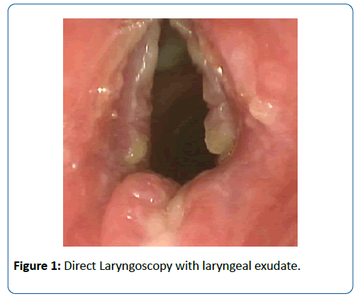
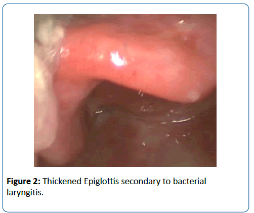
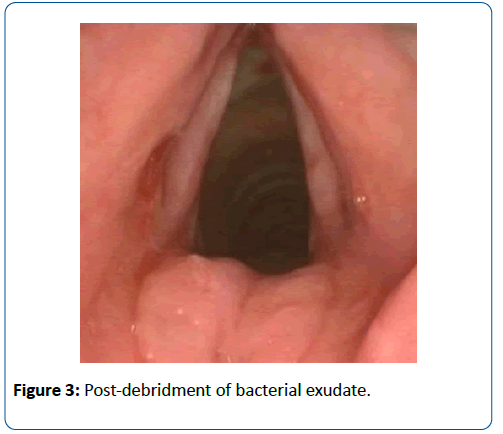
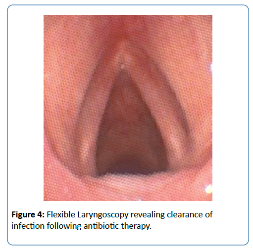
A 12-year-old Amish female with history of biliary atresia and hepatopulmonary syndrome presented for evaluation of persistent dysphonia and previous nasal fungal (Alternaria) infection on the day she was scheduled for liver transplant. Otolaryngology performed flexible laryngoscopy revealing supraglottic and glottic exudate and concern for infection. Direct Laryngoscopy with biopsy and culture revealed extensive colonization with MSSA and Strep G. Transplant was postponed and she was treated micafungin and vancomycin followed by nafcillin for one week. She was discharged on clindamycin and fluconazole therapy for four weeks. Repeat endoscopic examination 3 weeks after discharge showed complete resolution of the exudate (Figures 1-4) [1-3].
References
- Uslu C, Oysu C, Uklumen B (2008) Tuberculosis of the epiglottis: A case report. Eur Arch Otorhinolaryngol 265: 599-601.
- Williams RG, Tony DJ (1995) Mycobacterium marches back. J Laryngol Otol 109:5-13.
- Lin CJ, Kang BH, Wang HW (2002) Laryngeal tuberculosis masquerading as carcinoma. Eur Arch Otorhinolaryngol 259: 521-523.
Open Access Journals
- Aquaculture & Veterinary Science
- Chemistry & Chemical Sciences
- Clinical Sciences
- Engineering
- General Science
- Genetics & Molecular Biology
- Health Care & Nursing
- Immunology & Microbiology
- Materials Science
- Mathematics & Physics
- Medical Sciences
- Neurology & Psychiatry
- Oncology & Cancer Science
- Pharmaceutical Sciences
